

Immunology in the lab
19-November-2010
Immunology in the lab
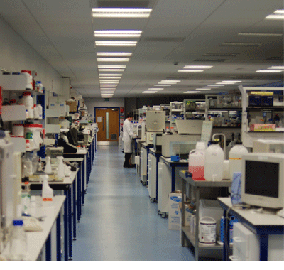
 When Jim Brewer pushes the heavy door open, the vista of gleaming glass and bright lights that meets our eyes seems to stretch to the horizon. There couldn't be a greater contrast, he says, between this lovely laboratory and the room he and Paul Garside shared twenty years ago, at the start of their Glasgow University careers.
"Imaging wasn't being done well in this field at that time," says Professor Brewer. "So I'd gone over on a fellowship to St Louis, where they had a great imaging centre, to learn all about it. Meanwhile Paul had gone to Minneapolis on a fellowship from the Wellcome Trust.
"When we came back there was no space left in the department. So they stuck us in this horrible lab in the basement of the old building, with big freezers full of blood samples in the middle.
"Naturally we talked to each other. Paul was looking at sections and I had been doing fluorescence work. We began to work together."
Perspex and superglue
Some years later both scientists began to see the potential for immunology of the new two-photon laser scanning microscopes. They began to apply for grants, says Professor Brewer. "At first we got a lot of 'You can't possibly do that'.
"So we had to plug away at it. We first got funding about seven years ago. We were lucky to have a creative young post-doc at the time. He loved building things out of perspex and superglue - which is just what you need at that stage of research with new equipment."
At this point the "Danger Laser" light winks out and he opens the door to the microscope room, where postdoctoral research fellow Robert Salmond has been working all day.
Once inside, Professor Brewer begins to explain about the microscope and the green and red moving images on the screen. "We are using the imaging system at the moment to look inside a lymph node. You have these in your armpits, around your groin, near your lungs, and around your neck and mouth.
On the frontier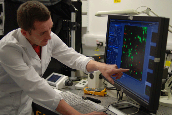
"Lymph nodes are like the forts of an army. The cells of the immune system are the soldiers. They circulate through the forts and constantly survey your whole body."
There are different types of soldiers in this army, just as there are in a real one, says Professor Brewer - each with its own job to do. "Inside the forts the soldiers that do the fighting talk to the scouts that are coming in from the frontier."
The green cells are the scouts, bringing news about a cut, a bite, a vaccine or a source of infection, he explains. The red cells are the fighters. "They're small now and don't look angry. But once activated they blow up and create a strong inflammatory response in that lymph node. That then grows larger and initiates an immune response."
The scientific name for the red soldiers is T-cells, Dr Salmond explains. "The green ones are dendritic cells. They have been exposed to a bite or infection and have moved to the lymph node to talk to the T-cells."
Illumination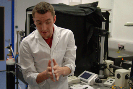
Something on the screen suddenly catches his eye. "Look at this," he says, turning to the professor, and they both watch closely as one of the scouts moves in on a small black patch on the screen.
"That looks like empty space," Robert explains. "But it's actually more cells."
Which demonstrates one big advantage of this microscope system, says Professor Brewer. "That tissue is opaque. But the microscope makes it seem transparent. With a conventional microscope you wouldn't see a thing."
Nice physics and engineering are what make the difference.
In conventional fluorescence microscopy a dye that fluoresces is added to the specimen. The parts that take up the dye then give off faint but visible light when stronger light is shone on them. This allows them to be studied through the microscope.
It's the same principle as in a fluorescent lamp. That gives off visible light from its fluorescent coating when hit by ultraviolet from the electrically excited gas in the bulb.
The key point about both fluorescent lamps and fluorescence microscopy is that the light coming out has a longer wavelength than the light going in. This is the big difference with the multi-photon microscope system.
Models
"What we do is hit the sample with two photons at once, using a high energy laser," says Professor Brewer. That means each photon needs only half the energy - and has twice the wavelength - of the single photon of light it then gives off. So to get visible light we can hit the sample with infrared.
 "The beauty of that is that infrared passes through tissue much more easily than visible light. So that is what makes our sample seem transparent. Infrared also carries less energy, so it's less damaging to the tissue."
This sophisticated system has opened the researchers' eyes to just how dynamic and complex the immune system is, says Professor Brewer. "It has also given us new insights in the fight against disease.
"But this is only the beginning. We're looking at two types of immune cell here, when there are maybe thirty. If we can learn about all the interactions between those, we could build a model of the whole system. Then we could test anything we wanted using that model.
He laughs. "That's the plan. By the time we've done all that I can retire."
Words used in pop-ups
| antigen | application | biomedical | complex | contract | design |
| device | electromagnetic radiation | electromagnetic spectrum | energy | experiment | focused |
| immune response | immune system | instrument | lymphatic system | magnifies | membrane |
| particle | pathogen | PhD | process | protein | protoplasm |
| radiation | research | researchers | stimulates | structure | subtle |
| thymus | tissue | ultraviolet | immunity | vessel | visible |
| X-rays |
For other websites and resources relevant to this science story try the